
An example of what can be revealed using my technology
This is an example to show what can be revealed using my technology, which quality level has been appreciated by experts virologists, microbiologists and biochemists.
Please check here what they had to say about it
I can start from an electron microscope image targeting 100nm objects and reveal way smaller objects in great details.
Here my input was a page from a publication, that was sent to me by a virologist, in order to put my technology at test.
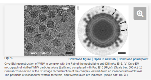
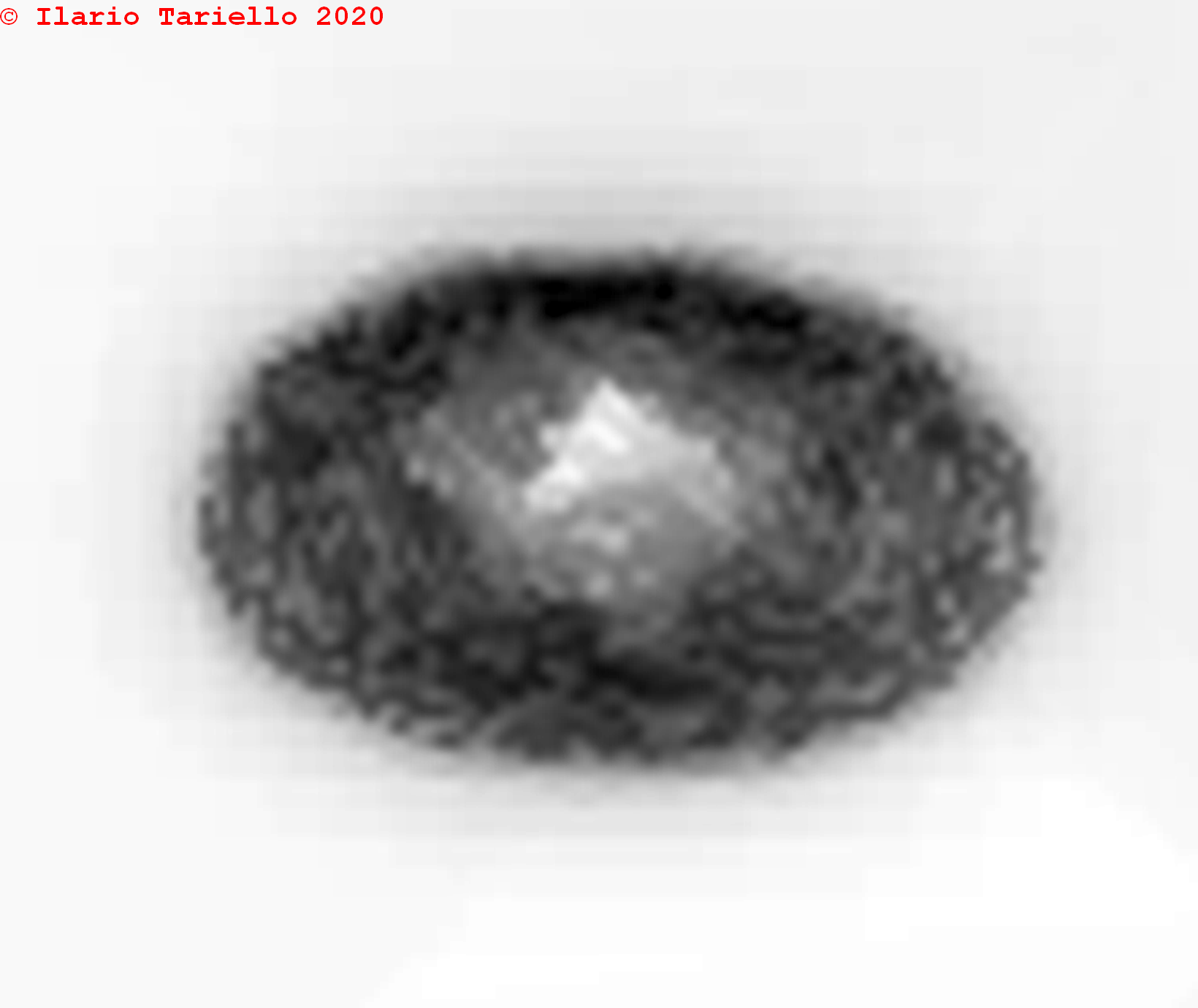
The page contained a picture of a bacteriophage (on the right side), a special virus eating bacteria, produced using an electron microscope.
The overall shape of the virion was clearly visible, but the component inside could not be distinguished.
I processed it in a few different ways and this is one of the first results I obtained.
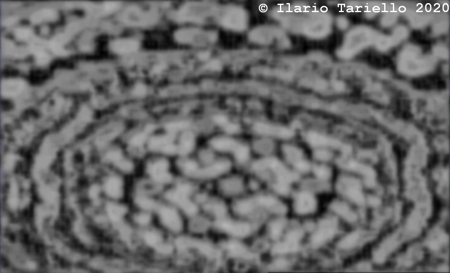
Here you can clearly see the single components inside its capsid, including the proteins, the genetic material and the lipidic membranes.
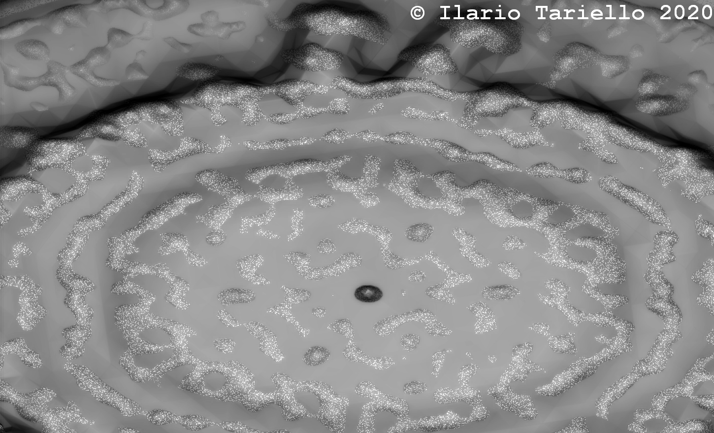
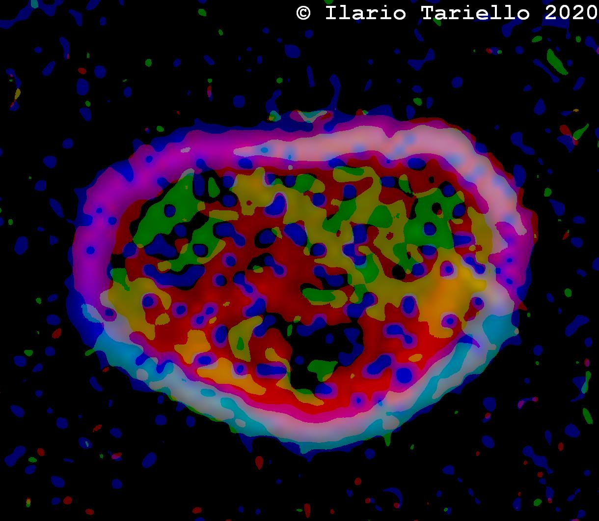
At this point I decided too dig deeper into it, targeting the very center of it, and I came out with several interesting pictures, among which for example the following ones.
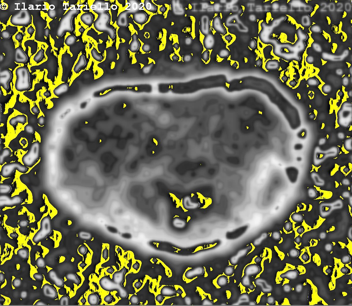
This is a different processing applied to the phage's picture.
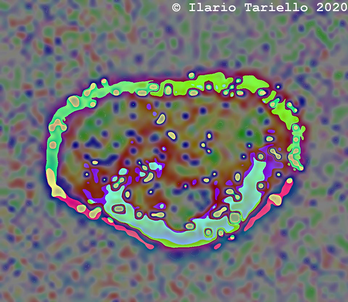
This one is a colorful resulting image of its center.
Please check here what they had to say about it
I can start from an electron microscope image targeting 100nm objects and reveal way smaller objects in great details.
Here my input was a page from a publication, that was sent to me by a virologist, in order to put my technology at test.

The page contained a picture of a bacteriophage (on the right side), a special virus eating bacteria, produced using an electron microscope.
The overall shape of the virion was clearly visible, but the component inside could not be distinguished.
I processed it in a few different ways and this is one of the first results I obtained.

Here you can clearly see the single components inside its capsid, including the proteins, the genetic material and the lipidic membranes.
At this point I decided too dig deeper into it, targeting the very center of it, and I came out with several interesting pictures, among which for example the following ones.


This is a different processing applied to the phage's picture.

This one is a colorful resulting image of its center.
